Adriana Dawes Summary: Research happening at the Dawes Lab
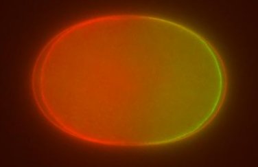
Single cell embryo of the nematode worm Caenorhabditis elegans
In the Dawes Lab, we are interested in understanding how biochemical pathways, mechanical structures, and cell geoemtry/morphology interact and regulate each other to produce the complex patterns we associate with biological systems. Misregulation of any of these aspects can have disastrous consequences ranging from birth defects to metastatic cancer. As an interdiscplinary lab, we use a wide variety of research techniques from mathematics, statistics, genetics and cell biology, and beyond. We develop biologically based theoretical models that are often both explanatory, identifying underlying mechanisms that give rise to seemingly contradictory behaviors, and predictive, providing novel direction for future experimental work. On the experimental side, we use the nematode worm Caenorhabditis elegans (C. elegans) as our experimental organism, exploiting the high level of conservation involved in these networks. Using an array of tools from genetics and microscopy, we perturb target proteins and quantify the resulting changes. Experimental data are used to motivate, challenge and expand our mathematical models. Tight integration and continuous feedback between experimental and theoretical results is critical to improving our understanding of these important regulatory networks and interactions.
Cell fate specification
Cells are exposed to many different signals in their external environment, and they must use those signals to make cell fate decisions, for instance whether to divide, grow or die. We are studying pathways that are critical in many developmental and disease processes: EGF/Ras and Notch. Theoretically, we have devised a biochemically based model consisting of coupled ODEs to simulate the response of a 6 cell array to spatially varying external signals. By separating parameter sets according to their simulated response to a drug treatment, we have used statistical techniques to determine which parts of the interaction network in the EGF and Notch pathways are responsible for species- specific differences. This project was initiated through the Research for Undergraduates: adventures in Mathematical Biology and its Applications (RUMBA) program and has so far involved 7 undergraduate students from both math and biology programs. The experimental portion of this project is led by Helen Chamberlin (OSU, Department of Molecular Genetics), where she has used an array of genetic and drug treatments to explore dose dependent differences in cell responses of members of the Caenorhabditis genus. We are experimentally characterizing the importance of the WNT pathway, a pathway component the model suggested was critical for the variable cell responses. We have also constructed and analyzed a simplified two variable model of the main interacting pathways to identify critical feedback loops and mechanisms for communication among the array of cells. We plan to construct a Boolean network model to more fully analyze the network architecture including its connectivity and sensitivity to noise.
Regulation of cortical actomyosin by Par and Rho proteins
The actoymosin cortex is a dynamic mechanical structure that gives rise to cell shape. The cortex is thought to be regulated by both Par and Rho protein families, although the mechanism of action is unknown. Par and Rho proteins are also able to spatially segregate within a single cell, creating two biochemically and physically distinct domains. Theoretically, we are using partial and stochastic differential equations to understand how changes in the abundance of proteins can give rise to the spatial patterns, and partial differential equations in a moving boundary simulation framework (with Ching-Shan Chou, OSU Department of Mathematics) to model changes in cortex shape in response to Par and Rho protein asymmetries. Experimentally, we use genetic perturbations such as RNA interference and the creation of new mutant strains to modulate the activity of specific proteins, and use confocal microscopy to quantify changes and reconstruct the likely network of interactions. With Dave Pilgrim, University of Alberta Department of Biological Sciences, we are also investigating the role of motor proteins in establishment and maintenance of protein asymmetries, and determining how different motor proteins may be acting cooperatively in key cellular processes such as cytokinesis and ring channel maintenance. We have been able to challenge the current dogma that Par and Rho proteins do not directly interact, and have uncovered fundamental mechanisms regulating cortical dynamics. Our future directions include developing an integrated and comprehensive modeling framework that can be used to study biochemical regulation of mechanical cortical dynamics in other biological contexts, and to biochemically characterize our proposed genetic interaction networks to determine whether the interactions are direct or indirect.
Force generation and pattern formation by microtubules
Microtubules are polymers that originate from centrosomes at the cell's nucleus and are responsible for properly segregating genetic material during division. The spindle, a structure made of microtubules, is positioned by force generating motor proteins located on the cortex, although the relationship between spindle position and force generation is not clear. Theoretically, we have developed and implemented a stochastic, individual based model to test hypotheses consistent with our experimental data (with Blerta Shtylla, Pomona College Department of Mathematics). Experimentally, we are optimizing a novel gene editing technique to insert pairs of fluorescent proteins that will act as a fluorescent timer to indicate if there are maturation specific differences in the centrosomes that could account for the observed spindle movement. We are developing a mean field model of spindle positioning in response to cortical force generation to to analyze the full range of potential behaviors and the effect of system parameters, and adapting quantitative imaging protocols to determine physical parameters of the spindle and force generating proteins.
Funding
We gratefully acknowledge funding from the following sources
- National Science Foundation
- National Institutes of Health
- Pelotonia
- Gordon and Betty Moore Foundation
Selected Publications
A full updated list of publications can be found [here.]