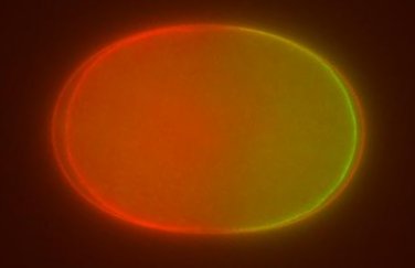
Single cell embryo of the nematode worm Caenorhabditis elegans
Cells polarize by segregating lipids, proteins and other molecules to discrete domains that are biochemically and physically distinct. Polarization is crucial for a wide variety of cellular processes including embryonic development, neurogenesis, directed motility and epithelial cell function. Many instances of polarization rely on a core group of proteins and lipids, including Rho proteins and Par proteins, as well as actin, myosin and tubulin. While cell types in many different organisms are capable of polarizing, single cell embryos of the nematode worm Caenorhabditis elegans have emerged as an important model system for the study of intracellular polarization. C. elegans requires polarization at the one cell stage for proper embryonic development and cell fate specification, and there are a number of well developed tools to facilitate research on polarization in this organism.
In the Dawes lab, we use a combination of theoretical and experimental approaches to study intracellular polarization in the early C. elegans embryo. We are investigating a number of questions regarding polarization, including the nature of the cue that initiates polarization, how mutual regulation of proteins and other factors establish and maintain polarized domains, and mechanisms that spatially position the boundary between domains. Theoretically, we use techniques from differential equations, dynamical systems and bifurcation theory to develop, simulate and analyze our mathematical models. Experimentally, we use fluorescent microscopy, RNA interference, and site directed mutagenesis to produce mutant organisms to challenge the results of our mathematical models.
Incorporating both theoretical and experimental approaches to investigate polarization allows us to uncover fundamental aspects of this conserved biological process. Feedback between theory and experiments allows us to better direct our experimental efforts and create more complete and accurate models.
Selected Publications
- White D, de Vries G, Martin J, Dawes AT (2015) Microtubule patterning in the presence of moving motor proteins. J Theor Biol 382:81-90. [PubMed link]
- Sturrock M, Dawes AT (2015) Protein abundance may regulate sensitivity to external cues in polarized cells. J Royal Soc, Interface 12(106). [PubMed link]
- Kravtsova N, Dawes AT (2014) Actomyosin regulation and symmetry breaking in a model of polarization in the early Caenorhabditis elegans embryo : symmetry breaking in cell polarization. Bull Math Biol 76(10):2426-48. [PubMed link]
- White D, de Vries G, Dawes AT (2014) Microtubule patterning in the presence of stationary motor distributions. Bull Math Biol 76(8):1917-40. [PubMed link]
- Dawes AT and Iron D (2013) Cortical geometry may influence placement of interface between Par protein domains in early Caenorhabditis elegans embryos. J Theor Biol May 9;333C:27-37. [PubMed link]
- Dawes AT and Munro EM (2011) PAR-3 oligomerization may provide an actin-independent mechanism to maintain distinct par protein domains in the early Caenorhabditis elegans embryo. Biophys J 101(6):1412-22. [PubMed link]
- Dawes AT (2009) A mathematical model of alpha-catenin dimerization at adherens junctions in polarized epithelial cells. J Theor Biol 257(3):480-8. [PubMed link]
- Dawes AT and Edelstein-Keshet L (2007) Phosphoinositides and Rho proteins spatially regulate actin polymerization to initiate and maintain directed movement in a one-dimensional model of a motile cell. Biophys J 92(3):744-68. [PubMed link]
- Maree AF, Jilkine A, Dawes AT, Grieneisen VA, Edelstein-Keshet L (2006) Polarization and movement of keratocytes: a multiscale modelling approach. Bull Math Biol 68(5):1169-211 [PubMed link]
- Dawes AT, Ermentrout GB, Cytrynbaum EN, Edelstein-Keshet L (2006) Actin filament branching and protrusion velocity in a simple 1D model of a motile cell. J Theor Biol 242(2):265-79. [PubMed link]
A full updated list of publications can be found [here.]